Escher’s Needlepoint
 Kaelyn
Male, Graduate Student, Cell Biology, Duke University
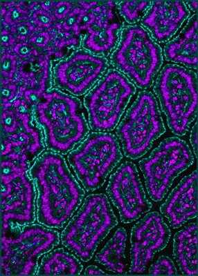
The surface of the gut is thrown into small projections (villi)
with invaginations
(crypts) at their base. The villi help to increase the area of
the gut surface for
absorption, while the crypts are the home of the stem cells that
are responsible
for continuous renewal of this epithelium. This is a
cross-section of villi and
crypts in the small intestine of an adult mouse. It is stained
to identify cell nuclei
(purple), and to show junctions between cells (green).
|
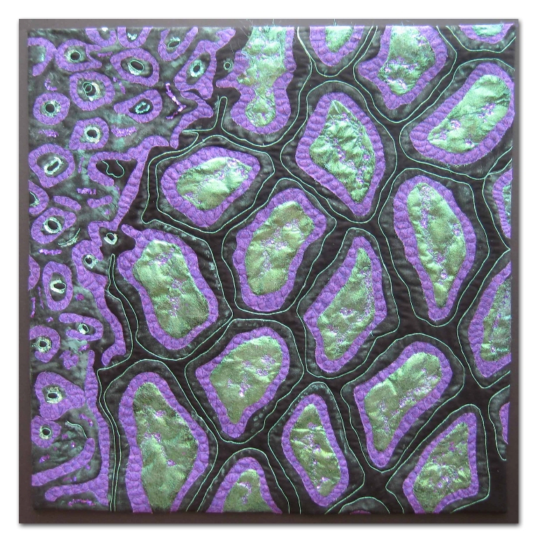
Lisa Ellis
I was immediately drawn to this photo by Kaelyn Male
when browsing the collection. I loved the colors and the
composition. Who knew that the gut could be so beautiful!
As a Mathematician, I am always drawn to Escher’s works
and could see why this piece was so named. I enjoyed playing
with embellishments to try to capture the sparkle of the green
and the three dimensional look of the purple. I used paint,
Angelina fibers and metallic bits for the inside of the cells.
Back to Gallery
|
 Fiber
Artists @ Loose Ends encourages members to explore new ideas and
techniques, inspires and nurtures creativity. By sharing our work
in private and public venues, we express our passion for the textile
medium.
Fiber
Artists @ Loose Ends encourages members to explore new ideas and
techniques, inspires and nurtures creativity. By sharing our work
in private and public venues, we express our passion for the textile
medium.