Loch Ness
 Åsa
Kolterud, Postdoctoral Research Fellow,
Cell & Developmental Biology, University of Michigan
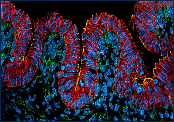
During embryogenesis, the developing intestine undergoes a
remarkable remodeling
process in which the surface of the gut tube is folded into
finger-like projections
called villi. These villi, which extend into the lumen of the
gut tube, drastically
increase the absorptive surface of the intestine and are
important for efficient
nutrient uptake. This photomicrograph shows the initial buckling
of the intestinal
epithelium (stained red) into nascent villi. The nuclei of the
cells are stained blue.
Note the flower-like nuclei within the epithelium – these are
dividing cells lining
up their chromosomes.
|

Carole Nicholas
The photograph of actively dividing intestinal cells, despite
its
title, does not conjure up an image of a deep, cold loch with
a mysterious, mythical monster lurking in its murky depths.
Rather, it looks to me like a joyous celebration, with a
colorful
storybook character Nessie emerging to engage in some magic
or whimsy. I used a Scottish highlands tartan for the binding
of the quilt. The background is moiré taffeta, to add texture
and movement. The cells are mostly dyed silk chiffon and
some cotton. Glass beads and metallic and rayon threads
were used as embellishment.
Back to Gallery
|
 Fiber
Artists @ Loose Ends encourages members to explore new ideas and
techniques, inspires and nurtures creativity. By sharing our work
in private and public venues, we express our passion for the textile
medium.
Fiber
Artists @ Loose Ends encourages members to explore new ideas and
techniques, inspires and nurtures creativity. By sharing our work
in private and public venues, we express our passion for the textile
medium.