Eureka
 Benjamin
Carlson, Graduate Student, Cell Biology, Duke University
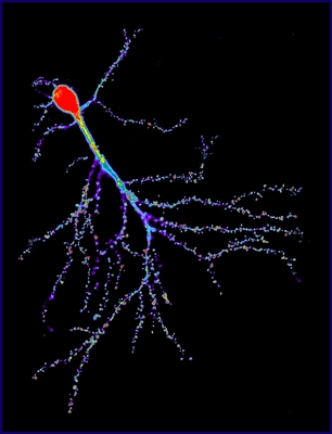
This image is of a mouse neuron from the hippocampus, the brain
area that
is critically involved in memory. The large branching
projections contain spiky
spines at their tips that receive signals from other neurons.
These spiny structures
are thought to be involved in the processing of information
during learning and
memory function. In this image, the neuron is stained to
identify a protein that
forms the internal skeleton of the cell. This allows us to study
the structure of
these spines during memory formation and learning.
|
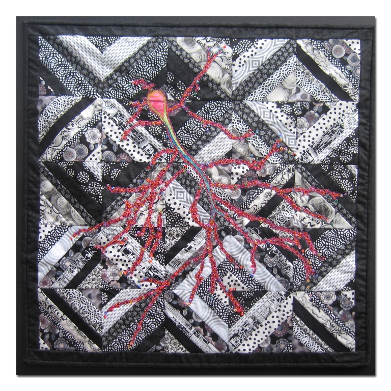
Donna DeSoto
I have to admit that the word “hippocampus” makes me smile!
When I first heard of the website containing BioArtography
images, I studied each one and read each description. As soon
as I found out that the subject of one of the images includes
the area of the brain that is involved in memory, I knew that
I wanted to make that quilt. The idea for the black and white
background of this piece came from Barbara Persing’s book
called StrataVarious Quilts. Materials used were commercial
cottons; the raw edge design was free motion quilted and glass
beads were applied by hand.
Back to Gallery
|
 Fiber
Artists @ Loose Ends encourages members to explore new ideas and
techniques, inspires and nurtures creativity. By sharing our work
in private and public venues, we express our passion for the textile
medium.
Fiber
Artists @ Loose Ends encourages members to explore new ideas and
techniques, inspires and nurtures creativity. By sharing our work
in private and public venues, we express our passion for the textile
medium.