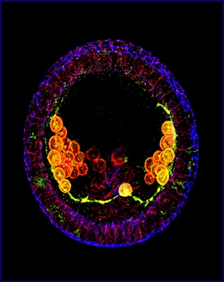
Crystal Ball
 Esther
Miranda, Research Analyst, Cell Biology, Duke University
Blue stains the outside of the urchin embryo almost like a cage
in a 3-D projection,
while the cells inside that will eventually form the skeleton
are stained red. The
green connecting fibers hold the cells together like clusters of
grapes, while the
bright yellow cells will form connective tissue. This complex
structure provides a
beautiful crystal ball for developmental studies.
|
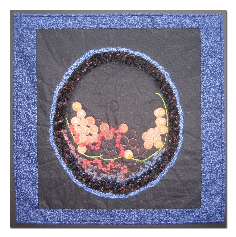
Donna DeSoto
Who knew that an urchin embryo could produce something as
dynamic as the photograph that was the basis for my artwork?
I love the colors and also the shapes and 3-D effect. I used a
number of cotton and synthetic fabrics and fibers, raw-edge
appliquéd, free-motion stitched and hand embellished. Photo
transfer was used to depict the cells that will form connective
tissue. This quilt is dedicated to the memory of my friend,
Patricia Botta.
Back to Gallery
|
 Fiber
Artists @ Loose Ends encourages members to explore new ideas and
techniques, inspires and nurtures creativity. By sharing our work
in private and public venues, we express our passion for the textile
medium.
Fiber
Artists @ Loose Ends encourages members to explore new ideas and
techniques, inspires and nurtures creativity. By sharing our work
in private and public venues, we express our passion for the textile
medium.